Sometimes there is a lump or swelling that appears on your pet.

This might be a

  • Dermal lump which is within the skin layer.
  • A subcutaneous lump which is under the skin layer.
  • An intra-abdominal lump which is in the abdomen.

The only way to know 100% what the lump is, is to remove it under general anaesthetic and send it to an external lab for analysis (histopathology). The pathologist will report what type of lump or tumour it is and if it has been completely removed or is likely to spread.

A less invasive procedure that may give us an indication of what sort of lump your pet has is to do a fine needle aspirate. 

The lump is secured.

A needle is placed into the centre of the lump.

The microscopic sample is expressed onto a microscope slide.

The slide is examined under the microscope.

The picture above shows a fatty mass called a lipoma. These are benign fatty lumps that grow slowly. The fat produces droplets seen microscopically often with little fat cells on closer examination.

An FNA is not 100% but gives your veterinarian an indication of what a lump may be and how it it likely to behave in the future. Any rapidly growing lump, painful lump or lump over 5cm in diameter is best excised and examined by a pathologist.

Other results may include

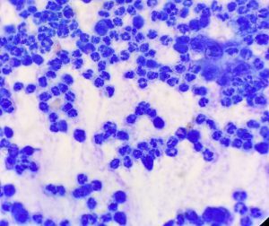
White Blood cells aspirated from an infected lump
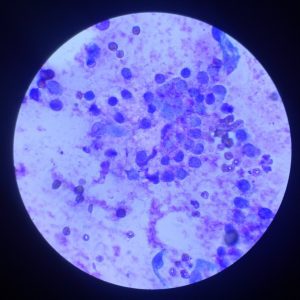
Tumours cells from a cutaneous lump. The granules in the cytoplasm indicate this may be a mast cell tumour.

If you are concerned about a lump or tumour on your pet. Book an appointment with one of our very experienced veterinarians to have it assessed.

  Make an Appointment with the Vet

or call us on 9913 7979


Register as a new client using our online form.

Are your pet's up to date with preventatives? Take our Preventative Health Review.

Google Rating
4.9
Based on 297 reviews
×
js_loader