X-ray is still one of the most valuable diagnostic tools of your veterinarian.
At Pittwater Animal Hospital there is a dedicated X-ray suite with recently updated digital radiography, adjustable radiographic table and dedicated computer consul. Images can be adjusted and magnified on our computer consul to examine for the smallest changes. All our xrays are then saved on the data base to refer back to in the future. If necessary images can be sent for further review.
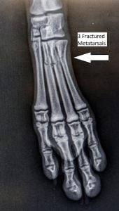
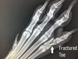
X-ray is a non-invasive procedure that may give information to your veterinarian about changes in bones or soft tissues of the animal. Some areas are more useful to x-ray such as the thoracic cavity (looking at the heart and lungs), bones and abdomen.
A 2 dimensional image is produced with the X-ray. To help with diagnosis, images from at least 2 different views, usually at perpendicular angles, are taken.
Possible x-rays performed at Pittwater Animal Hospital include
- Conscious x-ray during consultation
Sometimes during a consultation your veterinarian will wish an immediate image to guide treatment. Any breathing difficulties may prompt a chest x-ray. Possible intestinal blockage may lead to an abdominal x-ray. Possible fracture of a lower limb could encourage a conscious x-ray.
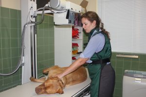
Conscious x-rays usually need the veterinarian to hold the patient, which increases the vets exposure to x-rays. It can be impossible to complete the procedure if animals are difficult to handle. It can also be challenging to take multiple views if required.
- X-ray under general anaesthetic
More comprehensive x-rays are best taken when our patients are under anaesthetic. This is particularly important for assessing bones such as elbows, shoulders, hips and knees. Positioning of the patient is key to taking the most useful image.
Most animals presenting for an x-ray under general anaesthetic, will be booked in for a surgery morning Monday to Friday. They will have a blood test prior to anaesthetic and will be on intravenous fluids throughout the procedure.
A full estimate will be given before your animal arrives for the procedure.


- Follow up x-ray
Changes over time can be very useful when monitoring your pet’s condition. Orthopaedic patients are often booked in for follow up x-rays 8 weeks after the procedure so the surgeon can monitor the position of implants and signs of healing. These images are usually taken either conscious or under sedation.
Often xrays will be taken in a series over time, especially if there is a concern that there may be an intestinal obstruction. A barium meal is sometimes given to patients with gut problems so we can outline the gut with this radio opaque liquid as it moves through the body.
Other follow up x-rays can be recommended if there are suspicious changes that may alter over time. This is very much the case with bone pathology or to monitor for the chance of an intestinal blockage. Scheduling follow up x-rays may avoid other more invasive steps in diagnosis.
Click Here to Make a Vet Appointment

